

What is an electrophysiology study?
An electrophysiology (EP) study is done to study the heart’s electrical activity. It is carried out by an electrophysiologist (a cardiologist specialising in the electrical system of the heart and heart rhythm disorders).
The heart pumps blood to the rest of the body through electrical signals. This signal originates from the sinoatrial node, the heart’s natural pacemaker, and is transmitted throughout the heart. This is responsible for the contractions of the heart. An EP study uses electrodes to detect these electrical signals and detect any kinds of fluctuations or abnormalities.
Why is an electrophysiology study done?
This study is done to detect any abnormalities in the heart. Your doctor can recommend this under the following conditions:
- You’re experiencing symptoms such as fainting, light-headedness, and weakness and your doctor suspects a cardiac cause behind it
- You have an abnormally fast, slow, or irregular heartbeat (arrhythmias)
- You have to get a pacemaker or defibrillator
- You have to undergo cardiac ablation
- You’re at an increased risk of sudden cardiac death
- Your doctor wants to check how well your heart medications are working to manage your condition
What is an arrhythmia?
Usually, the heart beats at 70-100 beats per minute. When the heartbeat is too fast, too slow, or irregular, it is called arrhythmia. It can occur in any age group, although the risk increases with age.
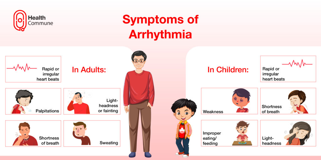
What are the symptoms of arrhythmia?
Arrhythmias can present a wide range of symptoms in both adults and children
In adults :
- Rapid or irregular heartbeat
- Palpitations
- Light-headedness or fainting
- Sweating
- Difficulty in breathing
In children :
- Rapid or irregular heartbeat
- Weakness
- Shortness of breath
- Light-headedness
- Improper eating/feeding
Are arrhythmias dangerous?
Arrhythmias can lead to an improper blood supply to the body. They can cause heart failure, loss of consciousness, sudden cardiac death, and stroke.
What is syncope? How is it related to heart disorders?
Syncope refers to a sudden loss of consciousness or fainting. It can have many causes.
If there is a heart disorder such as arrhythmia, blood and oxygen may not be delivered effectively to various parts of the body, including the brain. When this happens, it can lead to fainting.
How is an electrophysiology study done?
Can electrophysiology studies be done in children as well?
Yes, this can be performed on children as well.
What are the complications of an electrophysiology study?
Some complications include:
- Bleeding or infection at the catheter site
- Formation of a blood clot
- Damage to the vessel through which the catheter was inserted
- Damage to the conduction system of the heart
- Cardiac perforation (puncture to the wall of the heart)
How do I prepare for my electrophysiology study?
When your doctor thoroughly explains the procedure to you, you can express any concerns or questions you may have. Before the procedure, inform them about:
- Any medications (including vitamins, ayurvedic, herbal, and homoeopathy medicines) that you are taking, especially medicines like aspirin, and warfarin
- Any allergies, family history, or medical conditions that you have, especially disorders
- Any implants, piercings
You will be asked to fast overnight before the procedure. Remove your watch, jewellery , and any other metallic items that you have.
What happens during the procedure?
- You’ll be asked to change into the hospital gown
- The area where the catheter has to be inserted is shaved
- Your doctor establishes an IV line in your arm and connects you to an ECG
- You’ll receive a sedative and an anaesthetic, either a general one through a mask or at the site where the catheter is inserted
- Your doctor inserts the catheter into your blood vessel and views it from a screen (fluoroscopy)
- Once the catheter reaches the heart, your doctor sends minute electrical signals through the catheter to observe your heart function. This procedure, called cardiac mapping, is done to locate the area of the abnormal signals
- After the procedure is completed, the catheter is removed, and the entry site is tightly bandaged
What happens after the procedure?
- After the procedure, your healthcare providers monitor your vital signs and observe for any abnormalities
- You will receive pain medication for any kind of pain or discomfort
- You will be given specific instructions regarding :
- How to take care of the site
- What medications to take
- When to return to your daily routine
- What kind of activities to avoid
- Danger signs to watch out for, such as fever, bleeding, excessive pain or redness from the wound, light-headedness, fainting, and chest pain
What’s my next course of action?
After the procedure, your doctor will discuss your results with you, and based on this, you can be prescribed medications, or asked to undergo surgery.
