

AV malformation is a rare condition that can occur anywhere in the body. Know more about what it is, how it’s caused and its diagnosis and treatment.
What is an arteriovenous malformation (AVM)?
What happens in arteriovenous malformation?
Normally, blood flows from an artery, into the capillaries and then to the veins. This is important because the capillaries receive oxygen and nutrients from the artery and supply them to various tissues of the body. In the case of an arteriovenous malformation, blood flows directly from an artery into a vein, bypassing the capillary. As a result, the affected tissues receive less oxygen and nutrients.
Why do AVMs occur?
The exact cause is unknown. They are usually present from birth, but they grow and change with time. There’s an increased risk in certain conditions, like:
- Osler Weber Rendu syndrome (also called hereditary haemorrhagic telangiectasia), is a rare disease affecting the blood vessels in the body
- Cobb syndrome, a rare genetic disorder
- Parkes Weber syndrome, another rare disease with abnormal blood vessels throughout the body
What are the symptoms of arteriovenous malformation?
AVMs are often symptomless until they bleed. Depending on their location, they can have the following symptoms:
- In the brain:
- Headaches
- Confusion
- Weakness in one part of the body or loss of muscle strength
- Dizziness or fainting
- Nausea and vomiting
- Seizures
- Changes in vision
- Speech and language difficulties
- Changes in the sense of smell or taste
- Hearing loss
- Numbness, tingling or loss of sensation
- Hallucinations
- In the spinal cord:
- Back pain
- Weakness or loss of muscle strength
- In other locations:
- Lungs: Blood in cough, breathlessness
- Gastrointestinal tract: Abdominal pain, black stools
- Colour changes in your skin
- Sores or ulcers on your skin
What are the symptoms of AVM in children?
Your child may:
- Have a purple or bluish colour birthmark
- Complain of pain and warmth over the affected area
- Have pain or bleeding from the affected area
- Have a palpable pulse around the area
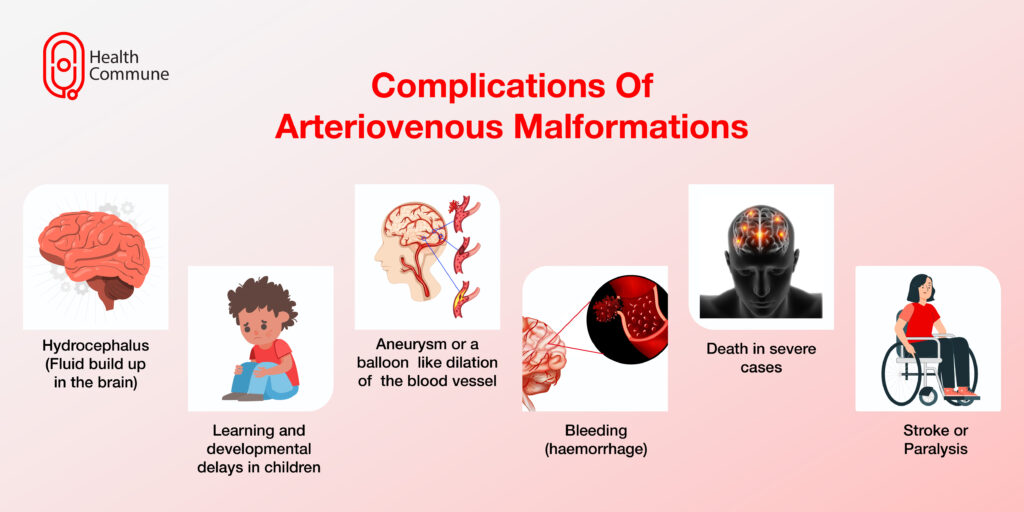
What are the complications of arteriovenous malformations?
Some complications include:
- Stroke
- Hydrocephalus (fluid build-up in the brain)
- Paralysis
- Bleeding (haemorrhage)
- Aneurysm, or a balloon-like dilatation of the blood vessel
- Learning and developmental delays in children
- Death, in very severe cases
How is an arteriovenous malformation diagnosed?
Your doctor will take your medical history and carry out a physical examination. They may try to listen to the blood flow in the suspected area. To confirm the diagnosis, they’ll prescribe some imaging tests:
- Ultrasound: An instrument called a transducer is pressed against the skin over the suspected area, which estimates the speed of the blood flow
- CT or MRI scans: These are employed to detect the exact location of the AVM and map the surrounding structures
- Computerised tomography (CT) angiogram: Through this procedure, your doctor can check if the blood flow is bypassing the capillaries. An iodine-rich contrast material is injected to check for the exact location of the malformation
- Magnetic resonance angiography (MRA): This is done to detect deeply located AVMs
How is an arteriovenous malformation treated?
Treatment depends on symptoms and the size and location of the AVM. Your doctor can prescribe medicines to control your pain and seizure episodes and to prevent bleeding. Surgery is the mainstay of treatment. These include:
- Endovascular embolisation: In this, a tube (catheter) is inserted through a blood vessel and guided to the AVM. A small coil, or stent, is then placed to redirect the blood flow
- Stereotactic surgery: This uses high-intensity radiation beams to stop the blood flow in the AV malformation
- Sclerotherapy: In this, a substance is injected into the arteriovenous malformation, which causes it to close. Scar tissue gets formed, which reduces the blood flow in the AVM
Is AVM preventable?
What’s the difference between an arteriovenous malformation and an arteriovenous fistula?
An AV malformation is usually present from birth, has multiple connections between the arteries and veins, is difficult to excise, and grows back frequently. In contrast, an AV fistula usually develops later on in life, has a single abnormal connection between the artery and the vein, and can be surgically excised with very low chances of recurrence.
